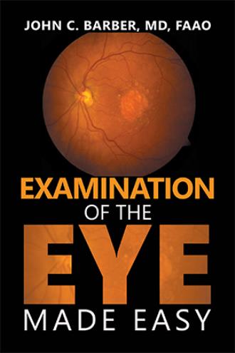
It is incumbent on every physician to be able to examine the eye and its adnexa. The eye may contain signs of many diseases that involve the entire body and the evidence needed to confirm the diagnosis that was suspected from the medical history and other parts of the physical examination of the body.
Because the eye contains nerves, vessels, muscles, pigmented tissues and connective tissues, derived from each of three embryonic layers, diseases that affect any of these tissues may cause changes in the eye. Both sensory and motor nerves have extensive roles in the movement and function of the eye.
The eye is the only place in the body where blood vessels can be seen and evaluated.
Patients with diabetes mellitus often have changes in the vessels of the retina as well as hemorrhages, exudates and infarcts within the retina. Diabetes is also the cause of palsies of the eye muscles and decrease in the visual acuity because of cataracts and retinal edema.
Hypertensive patients will have changes in the diameter of retinal arteries and pinching of the veins where they share a common muscular coat with arteries at vessel crossings. Hemorrhages within the retina from arterial hypertension have a characteristic shape and distribution.
Atherosclerosis of the arteries causes another characteristic change in the crossings of the blood vessels of the retina. Atherosclerosis causes the vessel walls to become opaque and obscure the blood in veins behind the arteries at Artery-Vein crossings resulting in a gap in the view of the venous blood that is wider that the bloodstream within the artery. The findings of hypertension and atherosclerosis may both be present simultaneously.
Forward protrusion of one or both eyes may be caused by malignant or benign tumors, bone changes, vascular anomalies, or thyroid disease prompting further examination and testing. Thyroid disease may affect any or all of the extraocular muscles.
Many other systemic diseases, i.e. lupus erythematosus, sarcoidosis, tuberculosis, fat emboli, and septicemia have specific findings within the eye, eyelids and orbit that can contribute to the correct diagnosis. Myasthenia gravis often presents with ptosis (drooping) of one eyelid or intermittent double vision related to weakness of extra-ocular muscles, but it must not be confused with partial paralysis of the third cranial nerve from diabetes or hypertension.
The findings within the eye may suggest a diagnosis of AIDS or ARCS that may lead to partial or total blindness if not discovered and treated before they become advanced.
The physician who does not examine the eye, both inside and out, does his patient and himself a major disservice.
The ease of using the direct ophthalmoscope to examine the retina depends to a large degree on the positioning of both the patient and the examiner. The preferred position is to have the patient seated on an elevated chair or an examination table, facing the examiner. The examiner stands or sits to the outside of the patient’s knees.
Alternatively, the patient may be examined lying on an exam table, looking at the ceiling. The examiner bends over the patient from the side to be examined.
The patient is asked to look at a specific spot, straight ahead, i.e. a clock, a picture, a piece of tape on the wall, etc. Telling the patient to look straight ahead will not make them hold the eye steady. Looking at a specific object will keep the eye still.
When examining the patient’s right eye, the examiner holds the ophthalmoscope in their right hand and looks through the aperture of the ophthalmoscope with the examiner’s right eye. Then use the left hand and left eye when examining the patient’s left eye. Unless the patient has a scarred cornea, a dense cataract or some other intraocular opacity, the light does not have to be at full power. There is a rheostat on the ophthalmoscope to adjust the light. For patient cooperation, it is best to use the minimum amount of light to produce a good image of the retina, while not temporarily blinding the patient.
The free hand is used to steady the patient and lift the upper eyelid. This is done by placing the fingers across the forehead above the eye and using the thumb to gently draw the loose skin of the eyelid up to the upper rim of the orbit. Gentle thumb pressure will hold the eye open without hurting the patient.
The examiner places the ophthalmoscope in front of their own eye when they are about ten inches from the patient. If possible, the examiner should keep both eyes open to avoid accommodation (focusing) with the examining eye. The ophthalmoscope is moved to direct the beam of light into the pupil. This should turn the pupil bright red. If the light is entering the pupil and the pupil is not red, there is a significant visual obstruction (corneal scar, hyphema, cataract, vitreous hemorrhage or vitreous membrane) within the eye and the retina may not be visible.
The examiner moves toward the patient, keeping the pupil centered in their vision. This will bring the eyes into alignment and the retina should come into view. If there is glare, the examiner should lower the ophthalmoscope slightly, i.e. usually less than 1 mm, until the glare is displaced away from the viewing aperture, so the retina is visible. Tilting the ophthalmoscope a degree or two down will also change the angle of the reflected light away from the viewing aperture. Some ophthalmoscopes have a polarized filter to reduce glare. This filter may not be sufficient to remove all glare if the above technique in not followed.
At this point the examiner is usually displaced temporally with respect to the optical axis of the patient. The patient should still be looking past the examiner at the spot on the wall, not at the examiner’s light, and may need to be reminded to look at the spot. This temporal displacement of the examiner will usually cause the examiner to be looking at the nasal retina, at or very close to the optic nerve head. The optic nerve head is the best place to start the examination. If the nerve head is not within the viewing area, use the vessels to find it. The branches of the vessels form Vs that point back along the vessels toward the nerve head.


